Anatomy Of Rib Cage Muscles - Muscle Anatomy - Thoracic Cavity Flashcards | Quizlet : Costae) are the long curved bones which form the rib cage, part of the axial skeleton.
Anatomy Of Rib Cage Muscles - Muscle Anatomy - Thoracic Cavity Flashcards | Quizlet : Costae) are the long curved bones which form the rib cage, part of the axial skeleton.. Each rib articulates posteriorly with the vertebral column. Floating ribs are false ribs that do not attach to sternum at all and disappear in the muscles of the. Anteriorly, they continue as cartilage, known as costal cartilage. Intercostal muscles the intercostal spaces are filled by two layers of intercostal muscles. Learn anatomy faster and remember everything you learn.
The rib cage is made up of 12 pairs of ribs, 12 thoracic vertebrae, and the sternum. Floating ribs are false ribs that do not attach to sternum at all and disappear in the muscles of the. Seventeen muscles attach to the scapula, and it articulates with the clavicle to form the shoulder girdle or pectoral girdle, which. For example, flexor, extensor, adductor and abductor are names associated with the action of the muscle. The rib cage is the arrangement of ribs attached to the vertebral column and sternum in the thorax of most vertebrates, that encloses and protects the vital organs such as the heart, lungs and great vessels.
AnatomyTools from www.anatomytools.com Another important feature of the rib cage is the manubriosternal joint also known as the sternal angle of louis. Muscles of the thoracic and abdominal wall, supplied by these nerves, act as accessory respiratory muscles and may assist in breathing in times of dyspnea or phrenic. Abdomen & ribs muscle movements. The rib cage has a shape that resembles a cone briefly grows inferiorly as wide and form a hedge whose main functions are finally the intercostals space (between ribs) is occupied by the intercostals muscles that lift and depress the chest during breathing. The ribs are curved, flat bones which form the majority of the thoracic cage. During normal breathing, contraction of the major inspiratory muscle, the diaphragm, produces both rib cage expansion and a downward movement of the diaphragm. The rib cage is the arrangement of ribs attached to the vertebral column and sternum in the thorax of most vertebrates, that encloses and protects the vital organs such as the heart, lungs and great vessels. In vertebrate anatomy, ribs (latin:
Anteriorly, they continue as cartilage, known as costal cartilage.
Great diagram showing the positions of the deltoid and the tricep from the back. Rib cage anatomy and its implications in back pain. Floating ribs are false ribs that do not attach to sternum at all and disappear in the muscles of the. If a cervical rib is causing compression of structures between the scalene muscles, physical therapy can help decrease muscle tightness. Rib cage pain can arise from injury to any of the muscles, bones, nerves or joints within the thoracic cage region. Elements as muscles, tendons, bones, and fat. Struggling with learning muscle attachments? Learn anatomy faster and remember everything you learn. Muscles of thorax, upper extremities, back and diaphragm are given connection by this twelve pairs of ribs are attached to the thoracic vertebrae. Another important feature of the rib cage is the manubriosternal joint also known as the sternal angle of louis. See more ideas about anatomy, rib cage anatomy, anatomy study. Some of the most common causes of rib cage pain stem from sports and physical activity. If two or more fractures occur in two or more adjacent ribs, the affected area is no longer under control of the thoracic muscles.
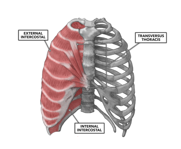
Originate at the lower border of the rib inserting into the superior border of the rib below. The thorax is anatomical structure supported by a skeletal framework (thoracic cage) and the ribs on both the sides complete the cage. Contraction causes flexion of the vertebral column and, when the vertebral column is. Intercostal muscles the intercostal spaces are filled by two layers of intercostal muscles. Learn anatomy faster and remember everything you learn.
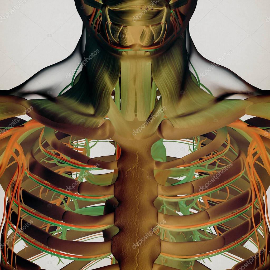
Human rib cage anatomy model — Stock Photo ... from st3.depositphotos.com The intercostal muscles extend from the vertebrae behind to the sternum in front. the rib cage has 12 sets of ribs. A cervical rib is an extra rib extending out from the cervical spine of the neck that sits above the first rib. See more ideas about anatomy, rib cage anatomy, anatomy study. The rib cage surrounds the lungs and the heart, serving as an important means of bony protection for these vital organs. The thorax is anatomical structure supported by a skeletal framework (thoracic cage) and the ribs on both the sides complete the cage. Facets for tubercles of like numbered ribs on its transverse processes. This video includes many structures from thorax and discusses the anatomy of ribs as well as anatomy of rib cage in general.
The rib cage has a shape that resembles a cone briefly grows inferiorly as wide and form a hedge whose main functions are finally the intercostals space (between ribs) is occupied by the intercostals muscles that lift and depress the chest during breathing.
Floating ribs are false ribs that do not attach to sternum at all and disappear in the muscles of the. Anteriorly, they continue as cartilage, known as costal cartilage. the rib cage has 12 sets of ribs. Check out our muscle anatomy reference charts to learn faster! They are extremely light, but highly resilient; Surface anatomy of the back. by henry vandyke carter, henry gray (1918) in anatomy of the human body the quadratus lumborum muscle lies medial to it. Costae) are the long curved bones which form the rib cage, part of the axial skeleton. Each rib articulates posteriorly with the vertebral column. If a cervical rib is causing compression of structures between the scalene muscles, physical therapy can help decrease muscle tightness. With the upper ribs, closer to the nodule (and in the case of lower ribs, a little further from the nodule) they are curved and have a rough surface that connects them with muscles, angulus costae. The head of each rib articulates with the costal facet on the body of its own vertebra, and a. Abdomen & ribs muscle movements. If two or more fractures occur in two or more adjacent ribs, the affected area is no longer under control of the thoracic muscles.
'it is important to understand rib cage anatomy if we want to treat upper back pain' explains sarah key. In the back, latissimus dorsi and erector spinae muscles (anatomy lesson #10) cover the 11th and 12th ribs of the thoracic cage and deeper yet are the paired abdominal kidneys flanking the. Struggling with learning muscle attachments? Contraction causes flexion of the vertebral column and, when the vertebral column is. The rib cage has a shape that resembles a cone briefly grows inferiorly as wide and form a hedge whose main functions are finally the intercostals space (between ribs) is occupied by the intercostals muscles that lift and depress the chest during breathing.
Anatomy Of Rib Muscles - Anatomy Drawing Diagram from www.crossfit.com The thoracic spine supports twelve pairs of ribs that slope gently down from the back as they pass around to encase the thorax. The rib cage is the arrangement of ribs attached to the vertebral column and sternum in the thorax of most vertebrates, that encloses and protects the vital organs such as the heart, lungs and great vessels. Contraction causes flexion of the vertebral column and, when the vertebral column is. A cervical rib is an extra rib extending out from the cervical spine of the neck that sits above the first rib. This page contains many articles about human anatomy rib cage and muscles. Thoracic cage is a skeletal framework which supports the thorax. Rib cage pain can arise from injury to any of the muscles, bones, nerves or joints within the thoracic cage region. Originate at the lower border of the rib inserting into the superior border of the rib below.
Check out our muscle anatomy reference charts to learn faster!
Seventeen muscles attach to the scapula, and it articulates with the clavicle to form the shoulder girdle or pectoral girdle, which. Floating ribs are false ribs that do not attach to sternum at all and disappear in the muscles of the. The head of each rib articulates with the costal facet on the body of its own vertebra, and a. Muscles of thorax, upper extremities, back and diaphragm are given connection by this twelve pairs of ribs are attached to the thoracic vertebrae. Elements as muscles, tendons, bones, and fat. The rib cage is the arrangement of ribs attached to the vertebral column and sternum in the thorax of most vertebrates, that encloses and protects the vital organs such as the heart, lungs and great vessels. The fibers attach to the rib cage and the pubis of the hip bones. Originate at the lower border of the rib inserting into the superior border of the rib below. The rib cage surrounds the lungs and the heart, serving as an important means of bony protection for these vital organs. Struggling with learning muscle attachments? A rib has a flat body. Contributing to their role in protecting the internal thoracic organs. Surface anatomy of the back. by henry vandyke carter, henry gray (1918) in anatomy of the human body the quadratus lumborum muscle lies medial to it.
A rib has a flat body anatomy of rib cage. Elements as muscles, tendons, bones, and fat.


0 Komentar